

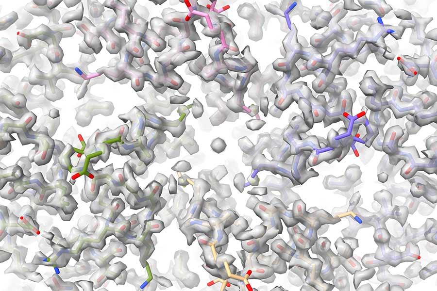
While technologies like X-rays allow us to create pictures of the inside of the human body, imaging on a molecular level is much more complicated. Cryogenic electron microscopy, or cryo-EM, allows scientists to reconstruct protein complexes, like viruses and antibodies, in three dimensions at nearly atomic-level resolutions. These reconstructions — which let scientists pinpoint the positions of individual atoms in a protein — can be used to understand the basis of diseases and design treatments to fight them.
Florida State University has been a key player in this specialized research since the field’s early days, and the university is now set to enhance the capabilities available both on campus and throughout the Southeast with construction of a new cryo-EM research center.
Researchers from FSU’s Department of Biological Science have received more than $5 million from the National Institutes of Health that will be used, in concert with additional university funding, to construct the Southeastern Center for Microscopy of MacroMolecular Machines, or the SECM4, at Florida State University.
Professors of Biological Science Scott Stagg and Kenneth Taylor will lead the service center in offering remote and in-person cryo-EM sample preparation, imaging, training and more to researchers at several universities in the area, making expensive cryo-EM instrumentation available to many more scientists who would otherwise not have access to this equipment.
Cryo-EM techniques come with a high cost, but they can yield highly rewarding outcomes: FSU faculty and students use cryo-EM to conduct research that directly affects human lives. Associate Professor of Biological Science Qian Yin, Professor Fanxiu Zhu and postdoctoral fellow Debipreeta Bhowmik recently published in the Proceedings of the National Academy of Science on their research uncovering the method some cancer-causing viruses use to avoid human immune system responses. Hamidreza Rahmani, a postdoctoral fellow working alongside Taylor, uses cryo-EM techniques to gain insight into the human heart and diseases that affect how it pumps blood to the body. Taylor and Rahmani’s research was published last year in PNAS.
“Accessibility is one of the biggest bottlenecks in the advancement of cryo-EM research as the field continues to expand,” said Stagg, who is also director of the FSU Biological Science Imaging Resource Facility, which supports additional types of microscopy work. “There are three other established centers in the country offering some data collection services, but we’re filling a niche in preparing, screening and optimizing samples to ensure their viability in creating an image. Researchers will also bring in their own samples, receive training and take the techniques back to their own labs.”
Traditional light microscopy works by bending light through a series of lenses in order to create a magnified image of something that would not otherwise be seen by the human eye. The wavelength of the light must be similar to the size of the sample being imaged, so traditional light microscopy can only image what is on the visual spectrum — what the human eye can see. Imaging atoms and proteins requires researchers to turn to electron microscopy, which uses electromagnets instead of lenses, and employs a beam of accelerated electrons as the source of illumination in order to image structures on an atomic level.
Diseases are often caused by mutations in proteins, and understanding the structure of these proteins is the first step in determining why the disease exists. By comparing mutated and non-mutated protein complexes, researchers can specify what in the structure causes the disease.
“With this new grant and the emerging center, FSU has solidified its place as a premier hub for cryo-electron microscopy in the Southeast,” said Sam Huckaba, dean of the College of Arts and Sciences. “I am so pleased to see this great success of our colleagues who have worked tirelessly to expand cryo-EM. Ken Taylor, who started us off years ago and brought this technique to FSU, and Scott Stagg are doing, with this recent funding success, outstanding work to enhance the world-class technical infrastructure we have at FSU that continues to draw researchers from across the country and the globe.”
Technologies at other FSU facilities, such as the protein expression and physical biochemistry suites in the Institute of Molecular Biophysics, the molecular cloning and sequencing facilities in the Department of Biological Science, and the physical biochemistry facility, will be utilized in conjunction with the new center to ensure the highest quality of sample preparation and analysis for researchers using the SECM4.
Some online services will become available by the end of the year, and the list of available services is projected to grow as construction on the SECM4 facility proceeds toward completion.