

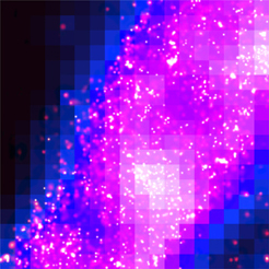
cell, as seen using the palm technique,
appears in violet.
A microscopy technique pioneered with the help of Florida State University’s National High Magnetic Field Laboratory has led to the development of a new light microscope capable of looking at proteins on a molecular level.
The new light microscope is so powerful it allows scientists to peer deep inside cells to see the fundamental organization of the key structures within. Developed by researchers at Howard Hughes Medical Institute’s Janelia Farm Research Campus in Virginia and the National Institutes of Health, in collaboration with FSU researchers Michael Davidson and Scott Olenych, the microscope is a boon to basic cell biology.
"As the technology advances, it may prove to be a key factor in unlocking the molecular-level secrets of intracellular dynamics," said Davidson, who directs the magnet lab’s Optical Microscopy Group.
The microscope and technology appear online in the Aug. 10 issue of Science Express.
The idea for the light microscope and the related new method, called photoactivated localization microscopy, or PALM, was conceived by physicists Eric Betzig and Harald Hess of the Howard Hughes Medical Institute, but they struggled with how to realize their vision. Awareness of biological tools being studied in Davidson’s lab ultimately inspired the two physicists’ plan to build a better microscope.
"In the world of biology, there is a new generation of fluorescent proteins that you can switch on at will with a little bit of violet light," Hess said. He and Betzig learned of these molecules, pioneered by Jennifer Lippincott-Schwartz and George Patterson at NIH, during conversations with Davidson.
Davidson suggested that these "optical highlighters" would be the best candidates for Betzig and Hess’ experiments. Davidson’s group then genetically engineered the highlighters and fused them to natural proteins in his lab. This technique allowed the researchers to attach a label to each copy of a protein they wished to study.
Here’s how the PALM technique works: The researchers label the molecules they want to study with a photoactivatable probe, and then expose those molecules to a small amount of violet light. The light activates fluorescence in a small percentage of molecules, and the microscope captures an image of those that are turned on until they bleach. The process is repeated approximately 10,000 times, with each repetition capturing the position of a different subset of molecules.
When a final image is created, it has a resolution previously only achievable with an electron microscope. However, the contrast in electron microscopy is more indiscriminate, whereas PALM can limit contrast to specific proteins of interest.
Lippincott-Schwartz said the use of PALM in conjunction with electron microscopy is particularly powerful.
"A great feature of PALM is that it can readily be used with electron microscopy, which produces a detailed image of very small structures – but not proteins – in cells," she said. "By correlating a PALM image showing protein distribution with an electron microscope image showing cell structure of the same sample, it becomes possible to understand how molecules are individually distributed in a cellular structure at the molecular scale."
The work was supported by the Howard Hughes Medical Institute and the National Institutes of Health.